|
Sac spider (Cheiracanthium) bites
back to Spider bites;
back to Cheiracanthium
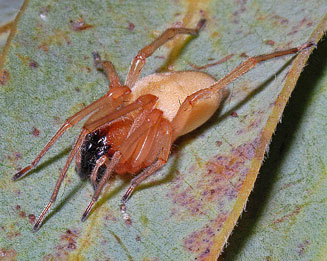
The Long-legged sac spider,
Cheiracanthium furculatum, previously
known as Cheiracanthium lawrencei, is synanthropic, entering houses, and
in South Africa has been implicated in 70-75% of spider bites that cause death
of tissue at the site of the bite (necrotic envenomations). No other
Cheiracanthium species are known to be implicated but should be regarded as of
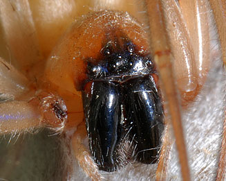
medical importance. Cheiracanthium furculatum has a black face and long anterior
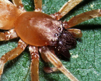
legs and must not be confused with the Leaf-curling sac spiders, Clubiona, of
the family Clubionidae, which do not have a black face and have the anterior and
posterior legs roughly equal in length. Clubiona is found away from
buildings.
 |
 |
|
Cheiracanthium furculatum. [image
N. Larsen ©] |
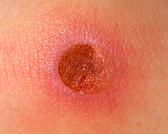
Wound from bite of Cheiracanthium furculatum.
[image N. Larsen ©] |
 |
 |
|
Head-on view of Cheiracanthium furculatum,
showing the black face. [image N. Larsen ©] |
Dorso-lateral view of Clubiona sp. It looks
similar to Cheiracanthium furculatum but does not have a black
face (although jaws are black). [image N. Larsen ©] |
Signs and symptoms
The bite of a Sac spider is at first painless or if noticed is accompanied by a
sharp burning pain, but as most bites occur at night, when the victim is asleep,
it is only noticed later with symptoms developing about 2-8 hours after the
bite. The bite presents as two tiny yellow-green spots 4-8mm apart where the
fangs penetrated the skin. These spots are the subcutaneous venom and with the
wide gape are diagnostic of Cheiracanthium envenomation; they are only visible during the
first day.
After 4-8 hours the area becomes inflamed (erythema, oedema and painful). A
bull’s eye lesion (lesion with a central haemorrhagic vesicle) may develop.
After 4-5 days the lesion sloughs leaving an irregular, round, ulcerated necrotic
wound, up to 10 mm in diameter. The wound is sterile, self limiting and starts to
heal spontaneously after 10 days but occasionally takes months, but in extreme cases
a skin graft is required. Some patients develop a mild fever, malaise and
headache after 1-3 days and the condition is sometimes misdiagnosed as tick bite
fever. However, tick bite fever symptoms develop after a 10 day incubation
period, by which stage the bite will have turned black and the surrounding area
swollen and red.
Differential Diagnosis
Sac spider bite symptoms could be confused with:
- tick-bite fever
- diabetic ulcers
- poor blood circulation
- allergic reactions
- Herpes simplex
- insect bites or stings
- viral or bacterial infections
- cellulitis
- necrotic anaerobic fasciitis
- bed and veld sores
Treatment
Treatment should be directed at the prevention of secondary infections. The bite
is sterile and the use of antibiotics is only required should infections set in
but this could be prevented by the use of an antibacterial cream and covered
with gauze to exclude secondary infection. No antivenom or
anti-tetanus injection is required. Spider venom contains an antibiotic thus
eliminating any infection.
Do not cut the site of the bite or use any traditional remedies as this
can aid the spread of
the venom and introduce infection. A doctor is usually only required if a secondary
infection occurs.
Publications (by date)
- Newlands G, Martindale C, Berson SD, Rippey JJ. 1980.
Cutaneous necrosis caused by the bite of Chiracanthium spiders. South African
Medical Journal 57: 171-173.
- Maretić Z. 1986. Spider venoms and their effect. In: Nentwig, W. (ed.),
Ecophysiology of spiders. Springer, New York, pp. 142-159.
- Newlands G. 1986. Necrotic arachnidism in southern Africa. Ph. D. thesis.
University of Witwatersrand, Johannesburg.
- Newlands G, Atkinson P. 1988. Review of southern African spiders of
medical importance, with notes on signs and symptoms of envenomation. South
African Medical Journal 73: 235-239.
- Newlands G. 1989. Anthropods that sting and bite man – their recognition
and treatment of patients. Journal of Continued Medical Education 17(7):
773-784.
- Newlands G, Atkinson P. 1990. A key for the clinical diagnosis of
araneism in Africa south of the equator. South African Medical Journal 77:
96-97.
- Filmer MR, Newlands G. 1994. Araneism in Africa south of the
equator with key to clinical diagnosis. Diseases of the Skin. 8(2): 4-10.
- Lotz LN. 1996. The genus Cheiracanthium (Araneae: Clubionidae) in southern
Africa. M. Sc. thesis. University of the Orange Free State, Bloemfontein, 1-96.
- Schrire L, Müller, GJ, Pantanowitz L. 1996. The diagnosis and
treatment of envenomation in South Africa. South African Institute for Medical
Research, Johannesburg, pp. 51.
- Croucamp W, Pantanowitz L. 1997. Necrotic araneism in South
Africa. Diseases of the skin. 11(5): 18-25.
- Müller GJ. 1999. Management of bites and stings: controversial
aspects. Abstract of the 6th African Arachnological Colloquium. African
Arachnological Society Newsletter 12.
- Croucamp W. 2000. Spider bites – diagnosis and management. Journal of
Continued Medical Education. 18(8): 670-678.
- Diaz JH. 2004. Global epidemiology, syndromic classification, management
and prevention of spider bites. American Journal of Tropical Medicine and
Hygiene. 71(2): 239–250.
- Müller GJ. 2005. Management of venomous bites and stings: A
mini-review. Unpublished notes.
- Snyman C, Larsen N. 2005. Spider bite and its treatment in southern
Africa. Occupational Health South Africa 11(2): 22-26.
- Lotz LN. 2007. The genus Cheiracanthium (Araneae: Miturgidae) in the
Afrotropical region. 1. Revision of known species. Navorsing van die Nasionale
Museum, Bloemfontein 10(1): 1-60.
Text by Norman Larsen © |