|
Spider Anatomy
Back to spider home
Arachnids have two body parts as opposed to insects with
three. The first part is the prosoma and the abdomen. usually referred to as the
cephalathorax, as it consists of a fused cephalic (head) and thorax which may be
separated by a distinct or indistinct cervical groove and the fovea.
The prosoma, usually referred to as the cephalathorax, as
it consists of a fused cephalic (head) and thorax which may be separated by a
distinct or indistinct cervical groove and the fovea, a depression to which the
stomach sucking muscles are attached. The shape of the fovea and cervical groove
are important diagnostic features for the identification of spiders into their
respective taxa. Chitin is responsible for this part of the exoskeleton being
hard and inflexible.
The prosoma houses various external appendages:
- A pair of chelicerae with hinged fangs
- A pair of pedipalps similar to the legs but with metatarsus absent. The
palps are often used as a pair of arms and in adult males the tarsus develop
secondary genitalia that give males the appearance of having boxing gloves.
These copulatory organs are vary important in the
identification of genera and species.
- Four pairs of legs consisting of seven segments.
apically is the coxa and trochanter (together forming the hip), the femur
(thigh), the petalla (knee), the tibia ( shin or lower leg), the metatarsus
(foot) and the tarsus (toes) with tarsal claws (toenails) at its end. The
legs are use for locomotion and prey capture.
- Dorsally the plate covering the prosoma is the
carapace which contains the simple eyes, up to eight in various positions,
an important identification character. The fovea a depression to
which the stomach sucking muscles are attached is an important diagnostic
feature for the identification of spiders into their respective taxa.
- Ventrally the endites (pedipalpal coxa) and the labium (lip) form the
mouth. The sternum (breast plate) with the dorsal carapace holds everything
together. the two are joined by a pleurae, an elastic membrane. It is along
the pleurae that the first crack stars the moulting process.
Internally the prosoma houses the central nervous system
(brain), venom glands, the sucking stomach with part of the intestine and the
muscles to control the appendages
The opisthosoma (abdomen), unlike the hard prosoma is soft
and pliable. this allows the abdomen to expand when saturated with food or to
house the developing eggs. Externally the abdomen contains:
- Booklungs (a primitive breathing apparatus) are situated
anterio-ventrally. Primitive mygalomorphae (baboon and trapdoor spiders)
have two pairs, Araneomorphae one pair and a tracheal spiracle (a breathing
apparatus as used by insects) and some advanced spiders have on tracheal
spiracles. the posterior edge of the booklungs is situated on the epigastric
furrow (fold) which house the spiders genitalia. Situated centrally on the
epigastric furrow the adult female has a blackish scleritised epigyne.
this epigyne is used in the identification of female spiders to generic and
species level.
- Posteriorly spiders have four to six spinnerets for the production of
silk.
- Above the spinnerets is the anal tubercle for the excretion waste
products.
The opisthosoma contains the respiratory organs, heart,
various spinning glands, the midgut, and ovaries for egg production.
|

|
|
External dorsal morphology of Araneomorpha |
 |
|
Left: External ventral view of Araneomorpha. Right: External ventral
view of Mygalomorpha. |
 |
|
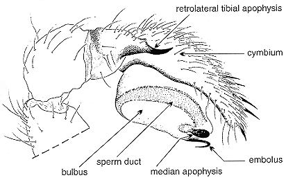
Copulatory organs: Male palp (entelegyne). lateral
view. |
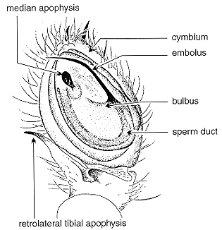 |
|
Copulatory organs: Male palp (entelegyne)
ventral view |
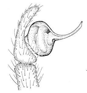 |
|
Copulatory organs: Male palp (haplogyne) |
|
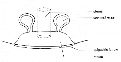
Copulatory organs: Female genitalia (haplogyne)
|
|
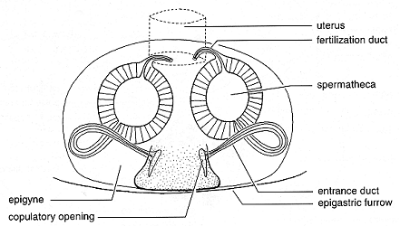
Copulatory organs: Female genitalia (entelegyne)
|
Illustrations from DIPPENAAR-SCHOEMAN, A. S. and JOCQUÉ,
R.1997. African Spiders. An Identification Manual. Plant Protection Research
Institute, Handbook No.9. 392 pp. Used with permission.
Text by Norman Larsen
© |